

What Part Of The Brain Controls Movement To The Arms And Legs
What is the brain?
The encephalon is a circuitous organ that controls thought, retentiveness, emotion, affect, motor skills, vision, breathing, temperature, hunger and every process that regulates our body. Together, the brain and spinal string that extends from it make upwardly the cardinal nervous system, or CNS.
What is the brain made of?
Weighing most iii pounds in the boilerplate adult, the brain is most threescore% fat. The remaining 40% is a combination of h2o, protein, carbohydrates and salts. The brain itself is a not a muscle. It contains blood vessels and nerves, including neurons and glial cells.
What is the gray matter and white affair?
Gray and white thing are ii unlike regions of the key nervous system. In the encephalon, gray matter refers to the darker, outer portion, while white thing describes the lighter, inner department underneath. In the spinal cord, this lodge is reversed: The white matter is on the outside, and the gray thing sits within.
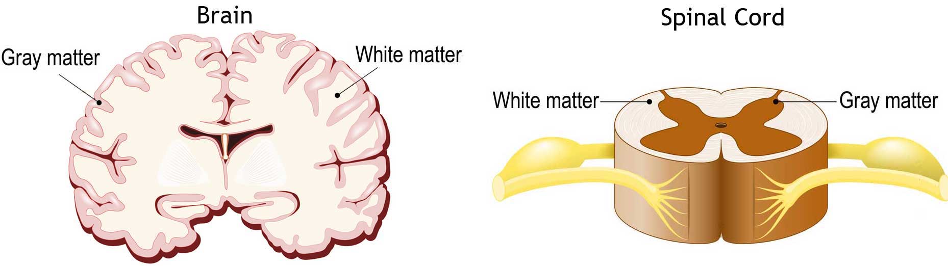
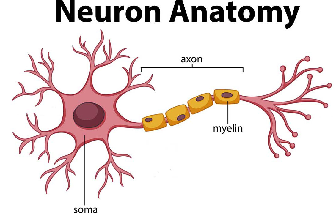
Gray thing is primarily composed of neuron somas (the round central cell bodies), and white matter is mostly made of axons (the long stems that connects neurons together) wrapped in myelin (a protective coating). The different composition of neuron parts is why the two appear as separate shades on certain scans.
Each region serves a different role. Gray matter is primarily responsible for processing and interpreting information, while white affair transmits that information to other parts of the nervous system.
How does the encephalon work?
The brain sends and receives chemical and electric signals throughout the trunk. Different signals command different processes, and your brain interprets each. Some make you feel tired, for example, while others make you feel pain.
Some messages are kept within the encephalon, while others are relayed through the spine and across the body'southward vast network of nerves to afar extremities. To practise this, the central nervous organization relies on billions of neurons (nerve cells).
Main Parts of the Brain and Their Functions
At a high level, the brain tin exist divided into the cerebrum, brainstem and cerebellum.
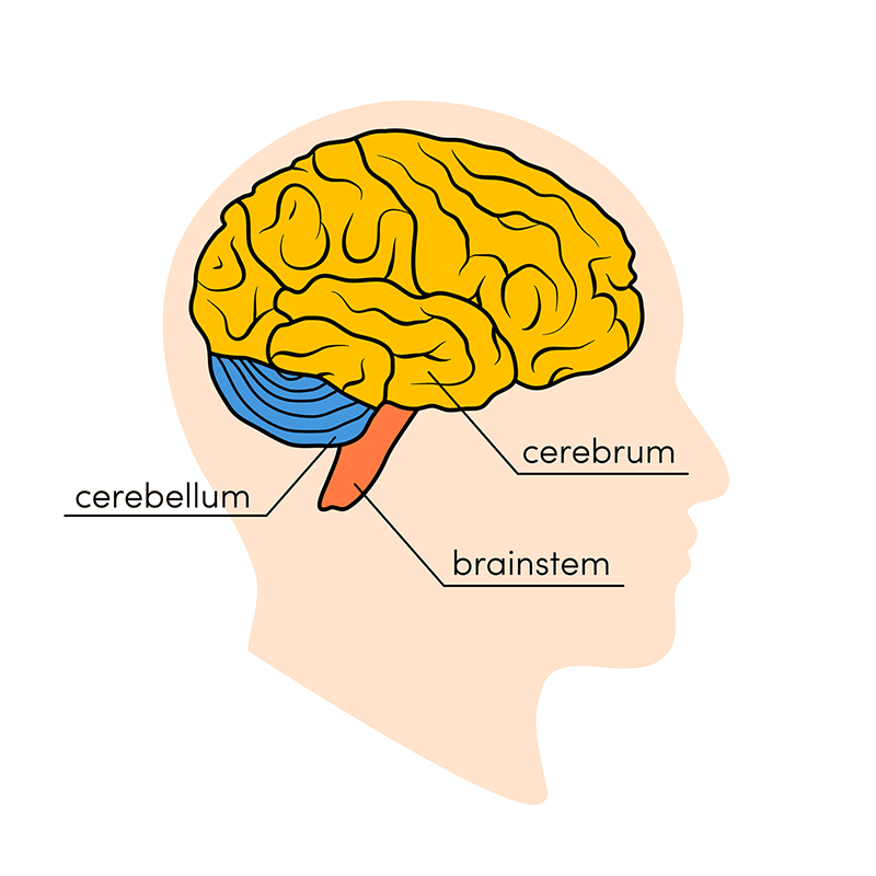
Cerebrum
The cerebrum (front end of encephalon) comprises gray matter (the cognitive cortex) and white affair at its middle. The largest part of the brain, the cerebrum initiates and coordinates movement and regulates temperature. Other areas of the cerebrum enable oral communication, judgment, thinking and reasoning, problem-solving, emotions and learning. Other functions relate to vision, hearing, touch and other senses.
Cerebral Cortex
Cortex is Latin for "bark," and describes the outer gray matter covering of the cerebrum. The cortex has a large surface expanse due to its folds, and comprises nearly one-half of the brain's weight.
The cerebral cortex is divided into ii halves, or hemispheres. Information technology is covered with ridges (gyri) and folds (sulci). The two halves join at a large, deep sulcus (the interhemispheric fissure, AKA the medial longitudinal fissure) that runs from the forepart of the caput to the dorsum. The right hemisphere controls the left side of the torso, and the left one-half controls the correct side of the body. The two halves communicate with i some other through a large, C-shaped construction of white matter and nerve pathways called the corpus callosum. The corpus callosum is in the center of the cerebrum.
Brainstem
The brainstem (middle of encephalon) connects the cerebrum with the spinal cord. The brainstem includes the midbrain, the pons and the medulla.
- Midbrain. The midbrain (or mesencephalon) is a very complex structure with a range of different neuron clusters (nuclei and colliculi), neural pathways and other structures. These features facilitate diverse functions, from hearing and movement to calculating responses and environmental changes. The midbrain too contains the substantia nigra, an area affected by Parkinson'southward disease that is rich in dopamine neurons and part of the basal ganglia, which enables movement and coordination.
- Pons. The pons is the origin for four of the 12 cranial nerves, which enable a range of activities such every bit tear product, chewing, blinking, focusing vision, balance, hearing and facial expression. Named for the Latin word for "bridge," the pons is the connection between the midbrain and the medulla.
- Medulla. At the bottom of the brainstem, the medulla is where the brain meets the spinal string. The medulla is essential to survival. Functions of the medulla regulate many bodily activities, including heart rhythm, breathing, blood flow, and oxygen and carbon dioxide levels. The medulla produces reflexive activities such equally sneezing, vomiting, coughing and swallowing.
The spinal cord extends from the bottom of the medulla and through a large opening in the bottom of the skull. Supported past the vertebrae, the spinal cord carries messages to and from the brain and the balance of the trunk.
Cerebellum
The cerebellum ("little brain") is a fist-sized portion of the brain located at the back of the head, beneath the temporal and occipital lobes and above the brainstem. Like the cognitive cortex, it has ii hemispheres. The outer portion contains neurons, and the inner area communicates with the cerebral cortex. Its function is to coordinate voluntary muscle movements and to maintain posture, balance and equilibrium. New studies are exploring the cerebellum's roles in thought, emotions and social behavior, equally well equally its possible involvement in addiction, autism and schizophrenia.
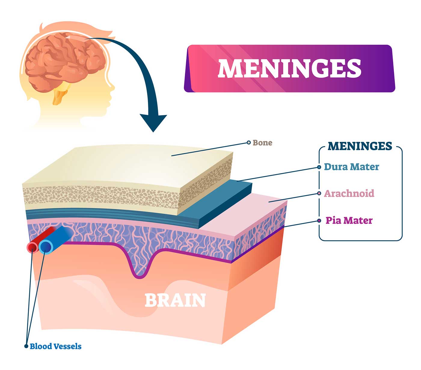
Brain Coverings: Meninges
Iii layers of protective covering called meninges surroundings the encephalon and the spinal string.
- The outermost layer, the dura mater, is thick and tough. It includes two layers: The periosteal layer of the dura mater lines the inner dome of the skull (cranium) and the meningeal layer is below that. Spaces between the layers allow for the passage of veins and arteries that supply blood menstruum to the brain.
- The arachnoid mater is a sparse, weblike layer of connective tissue that does not contain fretfulness or blood vessels. Below the arachnoid mater is the cerebrospinal fluid, or CSF. This fluid cushions the entire central nervous organisation (brain and spinal cord) and continually circulates around these structures to remove impurities.
- The pia mater is a thin membrane that hugs the surface of the brain and follows its contours. The pia mater is rich with veins and arteries.
Lobes of the Brain and What They Control
Each brain hemisphere (parts of the cerebrum) has iv sections, called lobes: frontal, parietal, temporal and occipital. Each lobe controls specific functions.
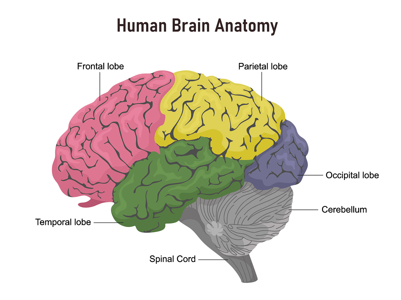
- Frontal lobe. The largest lobe of the encephalon, located in the front of the head, the frontal lobe is involved in personality characteristics, decision-making and movement. Recognition of olfactory property usually involves parts of the frontal lobe. The frontal lobe contains Broca'due south surface area, which is associated with speech power.
- Parietal lobe. The middle part of the brain, the parietal lobe helps a person place objects and understand spatial relationships (where one'southward trunk is compared with objects effectually the person). The parietal lobe is also involved in interpreting pain and impact in the body. The parietal lobe houses Wernicke's area, which helps the encephalon understand spoken linguistic communication.
- Occipital lobe. The occipital lobe is the back office of the brain that is involved with vision.
- Temporal lobe. The sides of the brain, temporal lobes are involved in curt-term memory, speech, musical rhythm and some degree of smell recognition.
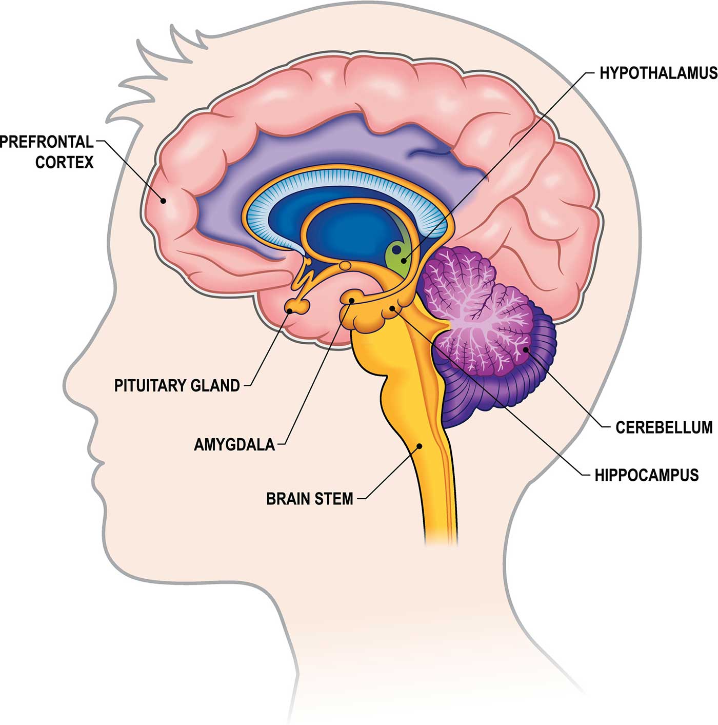
Deeper Structures Inside the Brain
Pituitary Gland
Sometimes called the "master gland," the pituitary gland is a pea-sized structure institute deep in the encephalon behind the bridge of the nose. The pituitary gland governs the office of other glands in the body, regulating the menstruum of hormones from the thyroid, adrenals, ovaries and testicles. It receives chemical signals from the hypothalamus through its stalk and claret supply.
Hypothalamus
The hypothalamus is located above the pituitary gland and sends it chemical messages that control its function. It regulates trunk temperature, synchronizes sleep patterns, controls hunger and thirst and likewise plays a role in some aspects of memory and emotion.
Amygdala
Pocket-sized, almond-shaped structures, an amygdala is located under each half (hemisphere) of the brain. Included in the limbic system, the amygdalae regulate emotion and retentiveness and are associated with the brain's reward arrangement, stress, and the "fight or flying" response when someone perceives a threat.
Hippocampus
A curved seahorse-shaped organ on the underside of each temporal lobe, the hippocampus is part of a larger structure called the hippocampal formation. It supports memory, learning, navigation and perception of space. Information technology receives information from the cerebral cortex and may play a role in Alzheimer'due south disease.
Pineal Gland
The pineal gland is located deep in the encephalon and attached by a stalk to the top of the third ventricle. The pineal gland responds to light and dark and secretes melatonin, which regulates circadian rhythms and the sleep-wake bike.
Ventricles and Cerebrospinal Fluid
Deep in the encephalon are 4 open up areas with passageways between them. They too open into the cardinal spinal canal and the area beneath arachnoid layer of the meninges.
The ventricles industry cerebrospinal fluid, or CSF, a watery fluid that circulates in and around the ventricles and the spinal cord, and between the meninges. CSF surrounds and cushions the spinal string and encephalon, washes out waste product and impurities, and delivers nutrients.
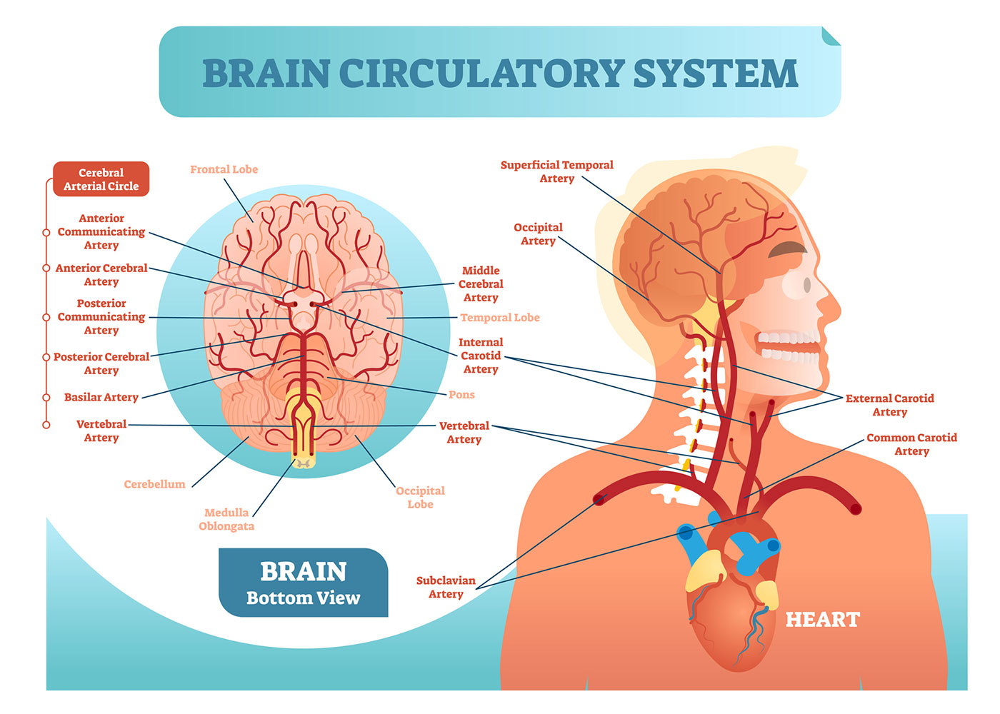
Blood Supply to the Brain
Two sets of claret vessels supply blood and oxygen to the brain: the vertebral arteries and the carotid arteries.
The external carotid arteries extend up the sides of your neck, and are where you tin can feel your pulse when y'all affect the area with your fingertips. The internal carotid arteries branch into the skull and circulate blood to the front part of the encephalon.
The vertebral arteries follow the spinal column into the skull, where they join together at the brainstem and form the basilar avenue, which supplies claret to the rear portions of the brain.
The circumvolve of Willis, a loop of claret vessels near the lesser of the brain that connects major arteries, circulates blood from the front of the encephalon to the back and helps the arterial systems communicate with one another.
Cranial Nerves
Inside the cranium (the dome of the skull), in that location are 12 fretfulness, chosen cranial nerves:
- Cranial nerve 1: The first is the olfactory nervus, which allows for your sense of smell.
- Cranial nerve 2: The optic nerve governs eyesight.
- Cranial nerve 3: The oculomotor nerve controls pupil response and other motions of the eye, and branches out from the surface area in the brainstem where the midbrain meets the pons.
- Cranial nerve 4: The trochlear nervus controls muscles in the eye. It emerges from the dorsum of the midbrain role of the brainstem.
- Cranial nerve v: The trigeminal nerve is the largest and most complex of the cranial fretfulness, with both sensory and motor function. It originates from the pons and conveys sensation from the scalp, teeth, jaw, sinuses, parts of the mouth and confront to the encephalon, allows the part of chewing muscles, and much more.
- Cranial nerve half-dozen: The abducens nerve innervates some of the muscles in the heart.
- Cranial nerve 7: The facial nerve supports face up move, taste, glandular and other functions.
- Cranial nervus eight: The vestibulocochlear nerve facilitates residue and hearing.
- Cranial nerve 9: The glossopharyngeal nervus allows taste, ear and throat movement, and has many more functions.
- Cranial nerve 10: The vagus nerve allows sensation around the ear and the digestive arrangement and controls motor activeness in the eye, throat and digestive organization.
- Cranial nerve xi: The accessory nerve innervates specific muscles in the head, cervix and shoulder.
- Cranial nerve 12: The hypoglossal nerve supplies motor activity to the tongue.
The first two nerves originate in the cerebrum, and the remaining 10 cranial nerves emerge from the brainstem, which has iii parts: the midbrain, the pons and the medulla.

What Part Of The Brain Controls Movement To The Arms And Legs,
Source: https://www.hopkinsmedicine.org/health/conditions-and-diseases/anatomy-of-the-brain
Posted by: leecouseed.blogspot.com
0 Response to "What Part Of The Brain Controls Movement To The Arms And Legs"
Post a Comment